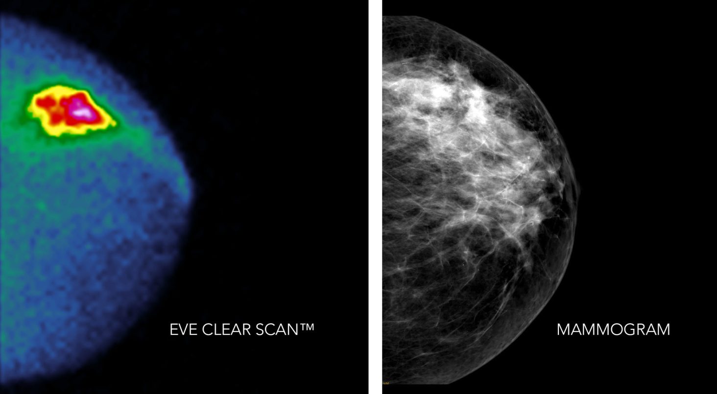
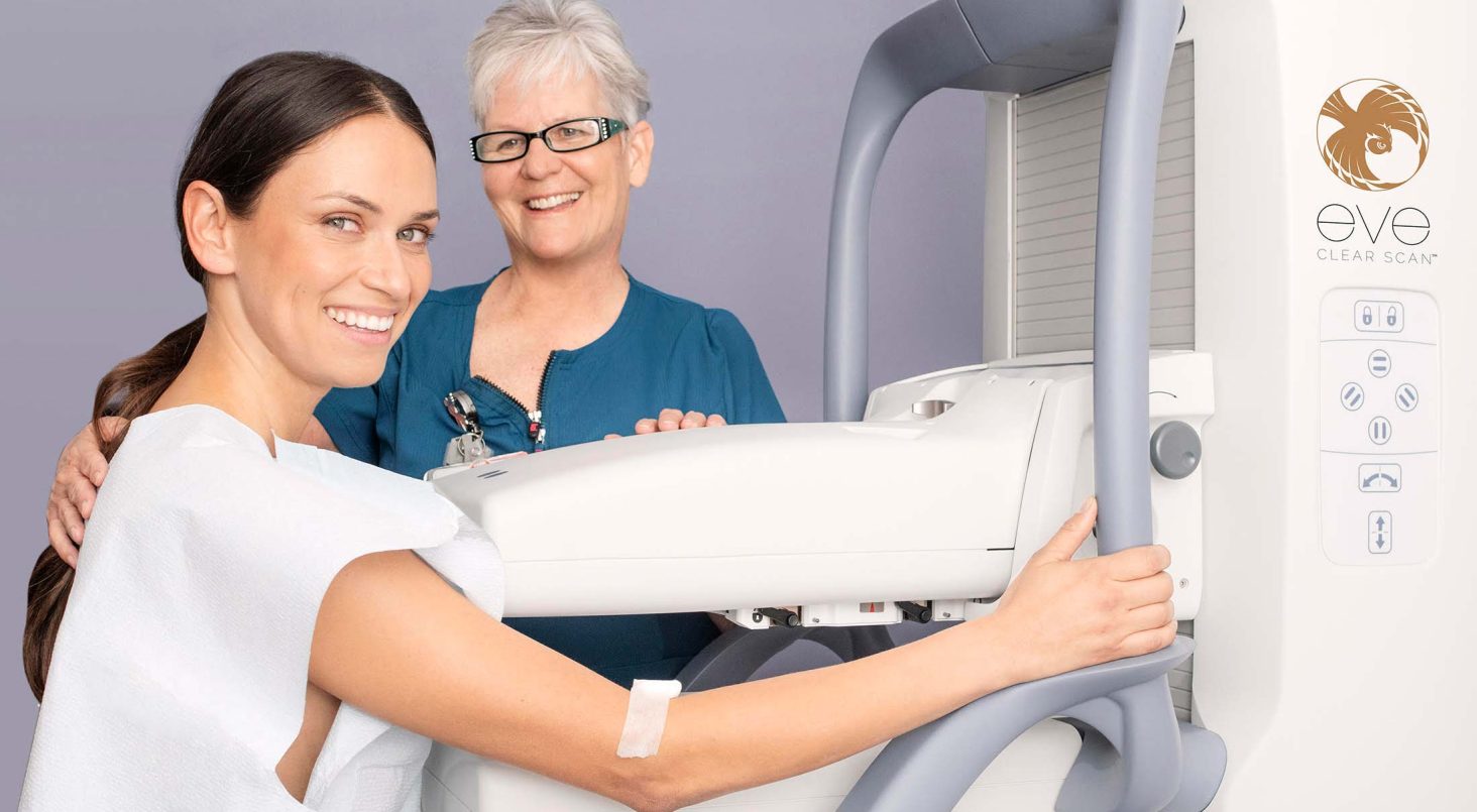
70%
of mammograms fail to depict early-stage cancer present in dense breasts
“Mammographic sensitivity... declined sharply... to 30% in women with extremely dense breasts.”*
*Mandelson, Ostreicher, et.al. Breast Density as a Predictor of Mammographic Detection: Comparison of Interval- and Screen-Detected Cancers. Journal of the National Cancer Institute, Volume 92, Issue 13, 5 July 2000, pp1081–1087.
Also Rhodes, Hruska, Connors, et.al. Molecular Breast Imaging at Reduced Radiation Dose for Supplemental Screening in Mammographically Dense Breasts. American Journal of Roentgenology. 2015 Feb. 204(2): 241-251.
3-4x
more early-stage cancers found by the MBI technology of SmartBreast compared to mammography. MBI also has fewer false positives (unnecessary biopsies).
“MBI is highly complementary to existing anatomical techniques, such as mammography, tomosynthesis and ultrasound.”*️
*O'Connor, Rhodes, Hruska. Molecular Breast Imaging. Expert Review of Anticancer Therapy. 2009 Aug; 9(8); 1072-1080
Also Michael O'Connor. Molecular breast imaging: an emerging modality for breast cancer screening. Breast Cancer Management.
Volume 4, No. 1. 7, 2015
44%
of additional MBI lesions were cancer (true positive) compared to only 28% for MRI in dense-breast patients being staged for surgery on newly found breast cancer. MRI resulted in 2x as many additional biopsies.
*Sumkin, Berg, Carter, et.al. Diagnostic Performance of MRI, Molecular Breast Imaging, and Contrast-enhanced Mammography in Women with Newly Diagnosed Breast Cancer. Radiology. 2019 Dec; 293(3): 531-540.
70%
of mammograms fail to depict early-stage cancer present in dense breasts
“Mammographic sensitivity... declined sharply... to 30% in women with extremely dense breasts.”*
*Mandelson, Ostreicher, et.al. Breast Density as a Predictor of Mammographic Detection: Comparison of Interval- and Screen-Detected Cancers. Journal of the National Cancer Institute, Volume 92, Issue 13, 5 July 2000, pp1081–1087.
Also Rhodes, Hruska, Connors, et.al. Molecular Breast Imaging at Reduced Radiation Dose for Supplemental Screening in Mammographically Dense Breasts. American Journal of Roentgenology. 2015 Feb. 204(2): 241-251.
3-4x
more early-stage cancers found by the MBI technology of Eve Clear Scan™ compared to mammography. MBI also has fewer false positives (unnecessary biopsies).
“MBI is highly complementary to existing anatomical techniques, such as mammography, tomosynthesis and ultrasound.”*️
*O'Connor, Rhodes, Hruska. Molecular Breast Imaging. Expert Review of Anticancer Therapy. 2009 Aug; 9(8); 1072-1080
Also Michael O'Connor. Molecular breast imaging: an emerging modality for breast cancer screening. Breast Cancer Management.
Volume 4, No. 1. 7, 2015
44%
of additional MBI lesions were cancer (true positive) compared to only 28% for MRI in dense-breast patients being staged for surgery on newly found breast cancer. MRI resulted in 2x as many additional biopsies.
*Sumkin, Berg, Carter, et.al. Diagnostic Performance of MRI, Molecular Breast Imaging, and Contrast-enhanced Mammography in Women with Newly Diagnosed Breast Cancer. Radiology. 2019 Dec; 293(3): 531-540.

Certainty

SmartBreast MBI provides a 3-4x increase in early stage cancer detection in dense breasts compared with 2D- and 3D- mammography.

Learn more

Clarity

SmartBreast's MBI technology depicts rapidly growing early-stage cancer cells, producing strikingly distinctive images in even the most dense breasts, often long before late-stage anatomic changes can be seen with mammography or MRI.

Learn more

Comfort

SmartBreast MBI's ergonomic design uses one or two cameras that immobilize the breast with gentle pressure, avoiding the crushing pain of mammography or the claustrophobia and noise of MRI.

Learn more

MBI Explained

MBI is a breast imaging technology that uses an intravenously injected cancer tracer and one or two gamma cameras to find areas of tracer uptake based on differences in cellular activity. Rapidly growing and dividing cells, such as cancer cells, take up the tracer and appear brighter than the less active healthy tissue. This is called "functional imaging" and is the SmartBreast difference that saves breasts and lives.

Learn more

What To Expect During An MBI With Eve Clear Scan

1

The cancer tracer is injected into the arm intravenously.

2

One breast is positioned between two gamma cameras (or between one gamma camera and a clear plastic plate) using minimal immobilizing pressure.

3

You remain still for a few minutes as an image is taken by the gamma camera(s).

4

Two images are usually taken for each breast, so you will be repositioned for each of the four images.

Resources On Dense Breast Tissue And Cancer

Are You Dense?

All about dense breasts. What is dense breast (DB) tissue? How do I know if I have DB tissue? What to do if I have DB tissue?

Learn more

Dense Breast Info

A resource for patients and providers. Topics like screening technologies and legislative information are discussed here.

Learn more

Susan G. Komen

A non-profit foundation that addresses a wide range of social and medical issues surrounding breast cancer
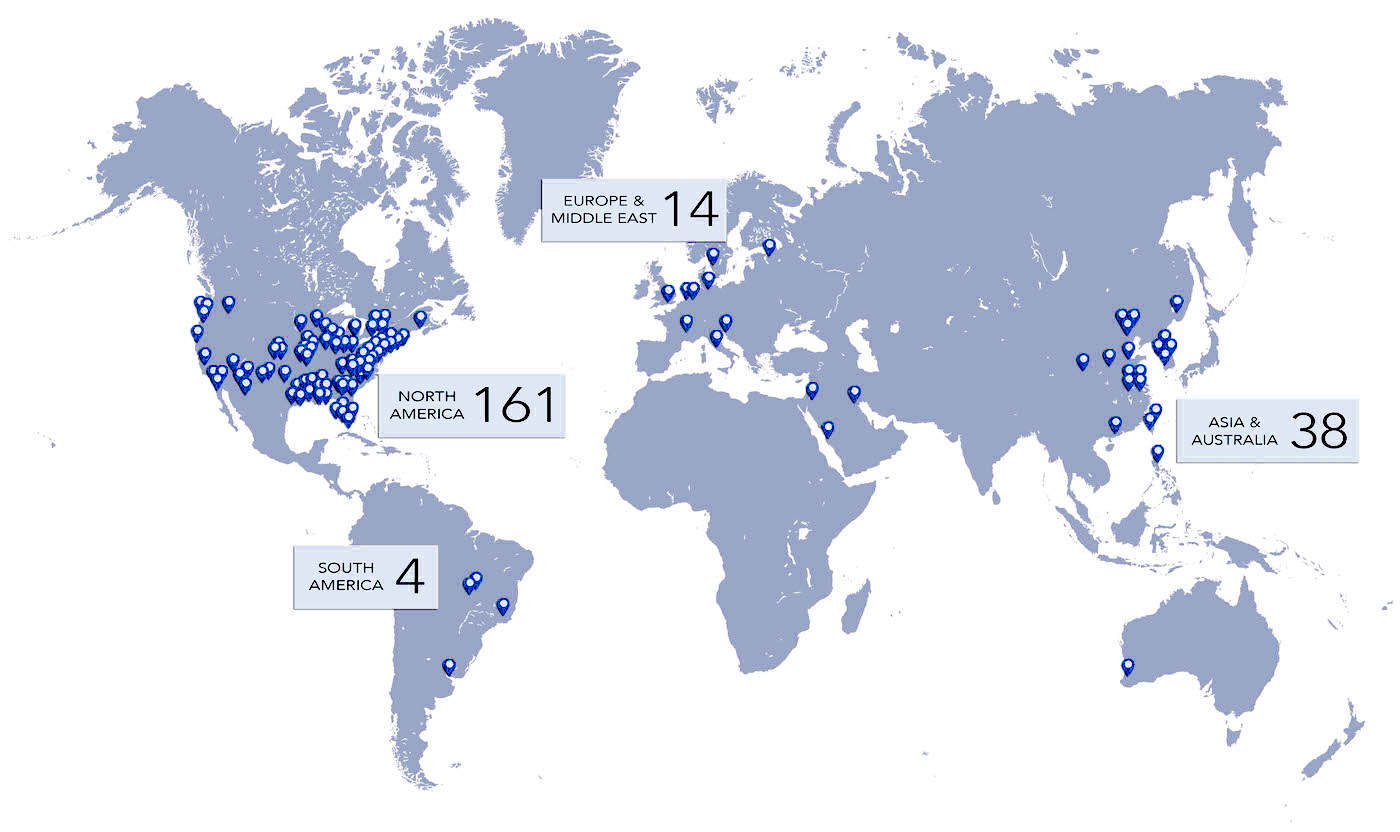
Learn more
Eve Clear Scan logo

Global Locations