Our 3C’s To Combat The Big C

Comfort
Death from breast cancer has significantly decreased since the advent of screening mammography.1-4 In addition to mortality reduction, early detection allows for a wider range of less invasive treatment options because early-stage cancer is easier to treat with very high success rates. Despite this benefit, up to 50% of women are deterred from having an annual screening mammogram because of pain experienced during the procedure. As many as 73% of women described mammography as mildly to severely painful.5
Pitfalls of Avoidance
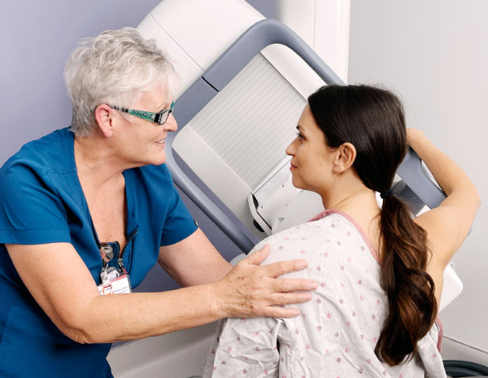
Mammography is performed with a patient standing in front of a special x-ray machine. A technologist places the breast on a detector plate. A plastic compression plate firmly presses the breast from above and flattens the breast while the X-ray is being taken. Flattening the breast is an essential requirement for mammography.
However, the pressure on the breast as it is secured between the two plates can generate pain. Studies have shown that several factors contribute to the pain:5-6
- sensitive breasts
- family history of breast diseases
- expectation of pain based on former mammography
- higher education
- anxiety
- breast sensitivity within the last three days
- insufficient care of the radiology technologist
Other factors like age, hormonal status, breast size, and hormone use were not associated with pain. It is worth noting that nearly half (46%) of women cited painful mammography as the cause of failure to repeat annual screening procedures.6
MBI: Staying in the Comfort Zone
To improve patient compliance in having a mammogram done, studies have looked into the measures that could mitigate procedural pain, primarily biological, psychological, and staff-related.7 After all, mammography still remains the generally accepted screening tool, and any measure to decrease procedural pain can result in improved patient compliance and early cancer detection.
Although the Eve Clear Scan MBI machine looks similar to a mammography machine, the core technologies are different, with MBI being used as a supplemental imaging modality in mammographically dense breast patients.

The Eve Clear Scan MBI Process
Step
1
Injection Of Gamma-Emitting Tracer
A gamma-emitting tracer (a contrast agent) will be injected into a vein in the arm. The tracer contains a substance that's quickly absorbed by fast-growing cancer cells. The gamma photons emitted by the tracer are detected by small gamma cameras that are part of the molecular breast imaging system.
Step
2
Positioning Of Breasts In Between Gamma Cameras
While seated in a chair facing the Eve Clear Scan MBI machine, the patient will be asked to open her gown and the technologist will place one breast on the flat surface of a gamma camera. The flat surface of a second gamma camera will be lowered gently on top of the breast, immobilizing it with a mild, non-painful compression, using only about 1/3rd of the force used in mammography (MMG) or digital breast tomosynthesis (DBT, also called “3D- mammography”).
Step
3
Imaging Procedure
The patient will sit still for a few minutes as the gamma cameras record the location of the tracer.
Step
4
Repositioning Of Breast For More Images
The breast will be repositioned for a second image. If both breasts are to be imaged, the other breast will be positioned in the imaging machine and the process will be repeated. Usually, a total of four images are recorded, just as in mammography screening. Women with larger breasts may need additional images.
References
1 Duffy SW, Tabar L, Chen HH, et al. “The impact of organized mammography service screening on breast carcinoma mortality in seven Swedish counties.” Cancer. 2002; 95(3): 458-469
2 Hellquist BN, Duffy SW, Abdsaleh S, et al. “Effectiveness of population-based service screening with mammography for women ages 40 to 49 years: evaluation of the Swedish Mammography Screening in Young Women (SCRY) cohort.” Cancer. 2011; 117(4): 714-722
3 Smart CR, Hendrick RE, Rutledge JH, 3rd, Smith RA. “Benefit of mammography screening in women ages 40 to 49 years. Current evidence from randomized controlled trials.” Cancer. 1995; 75(7): 1619-1626.
4 Tabar L, Vitak B, Chen TH, et al. Swedish two-county trial: impact of mammographic screening on breast cancer mortality during 3 decades. Radiology. 2011; 260(3): 658-663.
5 Keemers-Gels ME, Groenendijk RP, Van den Heuvel JH, et al. ”Pain experienced by women attending breast cancer screening.” Breast Cancer Res Treat. 2000 Apr; 60(3): 235-40.
6 Whelehan P, Evans A, Wells M, Macgillivray S. “The effect of mammography pain on repeat participation in breast cancer screening: a systematic review.” Breast. 2013 Aug; 22(4): 389-94.
7 Davey B. “Pain during mammography: Possible risk factors and ways to alleviate pain.” Radiology. Volume 13, Issue 3, 2007 Aug: 229-34.
